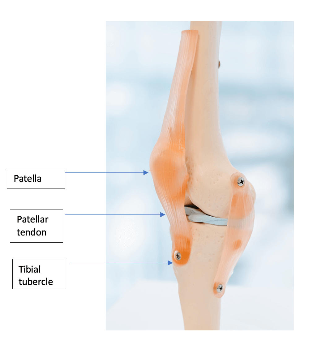
What is the tibial tubercle?
The tibial tubercle (or tibial tuberosity) is the lump on the front of your knee which is where the patellar tendon attaches from your patella (kneecap). It varies in shape and some people have a larger lump than others.
Why do I need a tibial tubercle osteotomy?
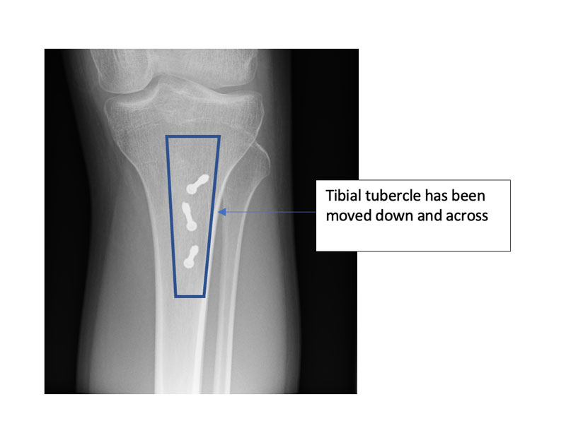
Changing the site of the attachment of the tibial tubercle can influence the position of the kneecap and make it more stable or offload a worn out area of cartilage to help with pain. Indications for a tibial tubercle osteotomy include lowering the kneecap if it is too high or moving it sideways. It can sometimes be used to help with knee pain in appropriate cases.
What is a tibial tubercle osteotomy?
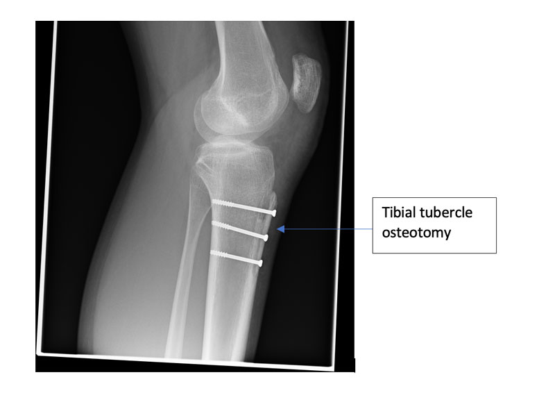
An osteotomy involves breaking and resetting a bone. In this case a tibial tubercle osteotomy involves using a saw to detach the tibial tuberosity and re-attach it with screws in a new position.
What are the possible complications of a tibial tubercle osteotomy?
All operations have risks and complications, some are general complications such as infection or clots in the veins (deep vein thrombosis). Others are more specific to the type of surgery.
The overall complication rate with this surgery is 5%
Some patients complain they can feel the screw heads when they kneel, if so the screws can be removed after a year from surgery as a day case procedure.
There is a 1% risk of the bones not healing, a 1% risk of fracture, 0.5% risk of pain, 0.5% risk of numbness and a 0.5% risk of infection.


What are the contra-indications to surgery?
If you smoke or have inflammation of your joints this may increase your risk of complications such as infection or the bone not healing which may mean this surgery is not appropriate for you. If you have a pain syndrome such as chronic regional pain syndrome (CRPS) you may be referred to a pain specialist in a pain clinic instead.
What happens after my surgery?
You will need to keep the wound dry for two weeks until the skin heals. You will be given some crutches to help you walk and help your balance. You will be allowed to put all your weight through the leg but you may not be allowed to bend it fully until the bone has healed. You will have a knee brace to protect the knee which allows you to bend it a little and this range of bend is increased until the bone has completely healed.
Regular X-rays are taken to monitor bone healing in the clinic and once we are satisfied that the bone has healed the brace is removed and your physiotherapist will work with you to increase your strength and balance.
